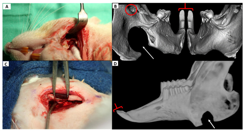
Figure 4.
Critical size defect (CSD) in the rat and rabbit alongside reconstructed µCT & CBCT images. In both animals, the defect is in the anterior ramus. (A): Surgical creation of the CSD in the rat. The metal retractor is reflecting the masseter and skin and the Q-tip is medializing the medial pterygoid to show the 5 mm defect. (B): A 3D CT reconstruction of the rat mandible from a caudal (posterior) vantage point 3 weeks following the surgery. The defect (thin arrow) remained fairly unchanged and symmetric incisor morphology (brackets) can be seen which was present at all time points. The circle indicates the mandibular foramen which transmits the inferior alveolar NVB. (C): Surgical creation of an 8 mm CSD in the anterior ramus of a rabbit with caliper shown. (D): A lateral 3D CT reconstruction of the same rabbit mandible 10 weeks later (the caudal view used in the rat was not suitable in the rabbit as it did effectively reveal relevant anatomy in a single view.) Note the CSD (thin arrow) is largely preserved as is symmetric incisor morphology (bracket) which was maintained at all measured time points. CSD: critical size defect, µCT= micro computed tomography, CBCT= cone beam computed tomography, NVB = neurovascular bundle.

