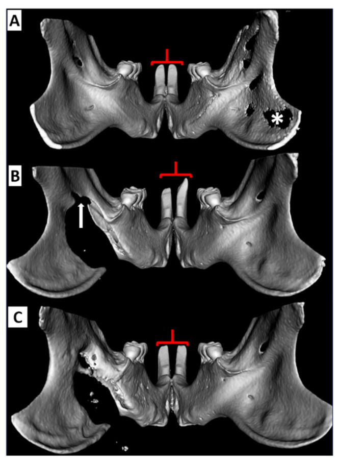
Figure 5.
Reconstructed µCT images showing effects of ligation of the left inferior alveolar NVB & ID in the rat incisors: (A) Baseline image prior to defect creation. Note the incisor symmetry (red brackets). Volume averaging of the thin ramus bone on the right results in the artifactual appearance of no bone (*). (B) One week following defect creation and ligation of the NVB (arrow). Note in this case that there is bilateral incisor dysmorphology but the incisor contralateral (to the defect) is considerably longer. (C) Three weeks following defect creation there is complete restoration of incisor length and morphology. CBCT = cone beam computed tomography, µCT = micro computed tomography NVB: neurovascular bundle, ID: intermediate defect.

