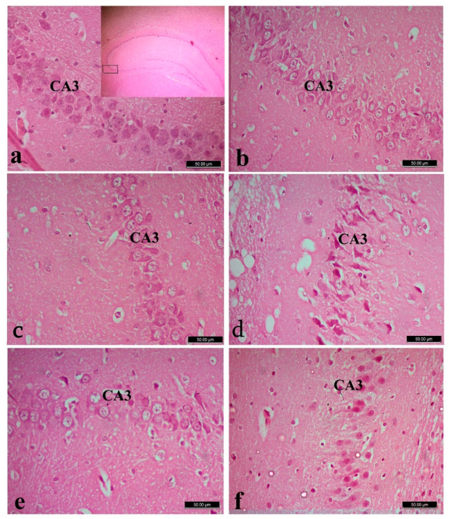
Figure 3.
Hippocampus of control (a) with right pan window showed the area of CA3 in hippocampus, L-arginine (b), L-carnitine (c) and fipronil (FPN)-treated rats (d). FPN-treated rat showed decreased thickness and degeneration of pyramidal cell layer in the CA3 region, with dystrophic changes in the form of shrunken hyperchromatic, with irregular distribution and degenerated neurocytes. Improvement is observed in FPN + L-arginine and FPN + L-carnitine-treated groups (e,f). Stain: Hematoxylin and Eosin (H&E), magnification 400×.

