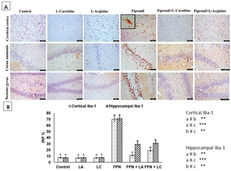
Figure 5.
(A) Immunohistochemical staining of the cerebral cortex, CA region and dentate gyrus with Iba-1. Negative and non-observed immunostaining were seen in control, L-arginine (LA) and L-carnitine (LC) groups. Fipronil (FPN) group showed positive brownish Iba-1 immunoreactive microglia with numerous fine branching processes nuclei. Reduced immunoreactivity of microglia in FPN + LA and FPN + LC treated groups was seen [Anti-Iba-1 × 400]. (B) Immunoreactive parts percentage (IRP%) of Iba-1 protein expressed as mean ± SE. Symbols **, *** indicates significant p value < 0.01 and 0.001, respectively.

