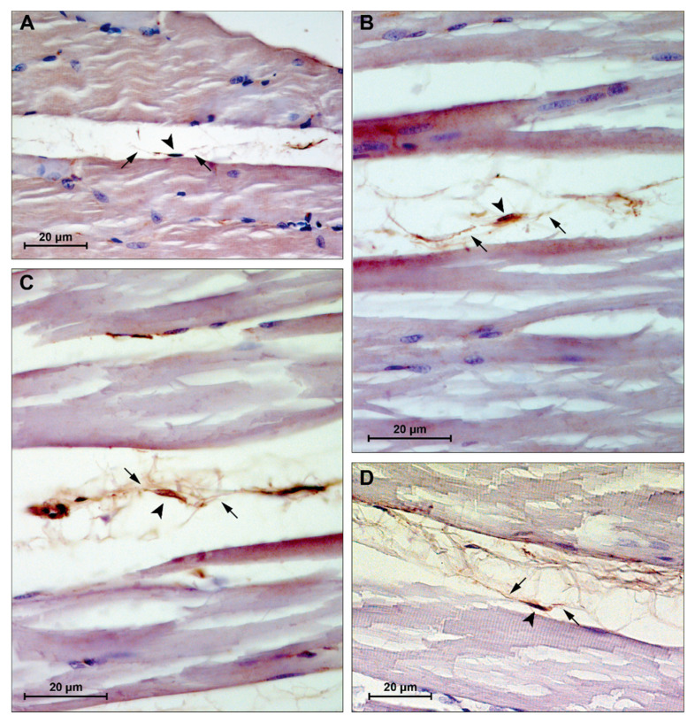
Figure 7.
Representative images of immunohistochemistry staining for muscle tissue CD34+ cells. (A) group CTRL4W, estimated size body (arrowhead): 5.23 μm, estimated cytoplasmic processes length (arrows): 13.28 and 24.28 μm; (B) group PA4W, estimated size body (arrowhead): 5.04 μm, estimated cytoplasmic processes length (arrows): 26.68 and 11.94 μm; (C) group CTRL16W, estimated size body (arrowhead): 8.69 μm, estimated cytoplasmic processes length (arrows): 13.07 and 20.56 μm; (D) group PA16W, estimated size body (arrowhead): 9.56 μm, estimated cytoplasmic processes length (arrows): 22.03 and 13.36 μm. Lens magnification: ×40. Scale bars: 20 μm. CD34+ cells nuclei and cytoplasmic processes were measured using a caliper tool of the software for image acquisition (AxioVision Release 4.8.2—SP2 Software, Carl Zeiss Microscopy GmbH, Jena, Germany). CTRL4W, control sedentary rats sacrificed at 4 weeks; PA4W, rats performing physical exercise sacrificed at 4 weeks; CTRL16W, control sedentary rats sacrificed at 16 weeks; PA16W, rats performing physical exercise sacrificed at 4 weeks.

