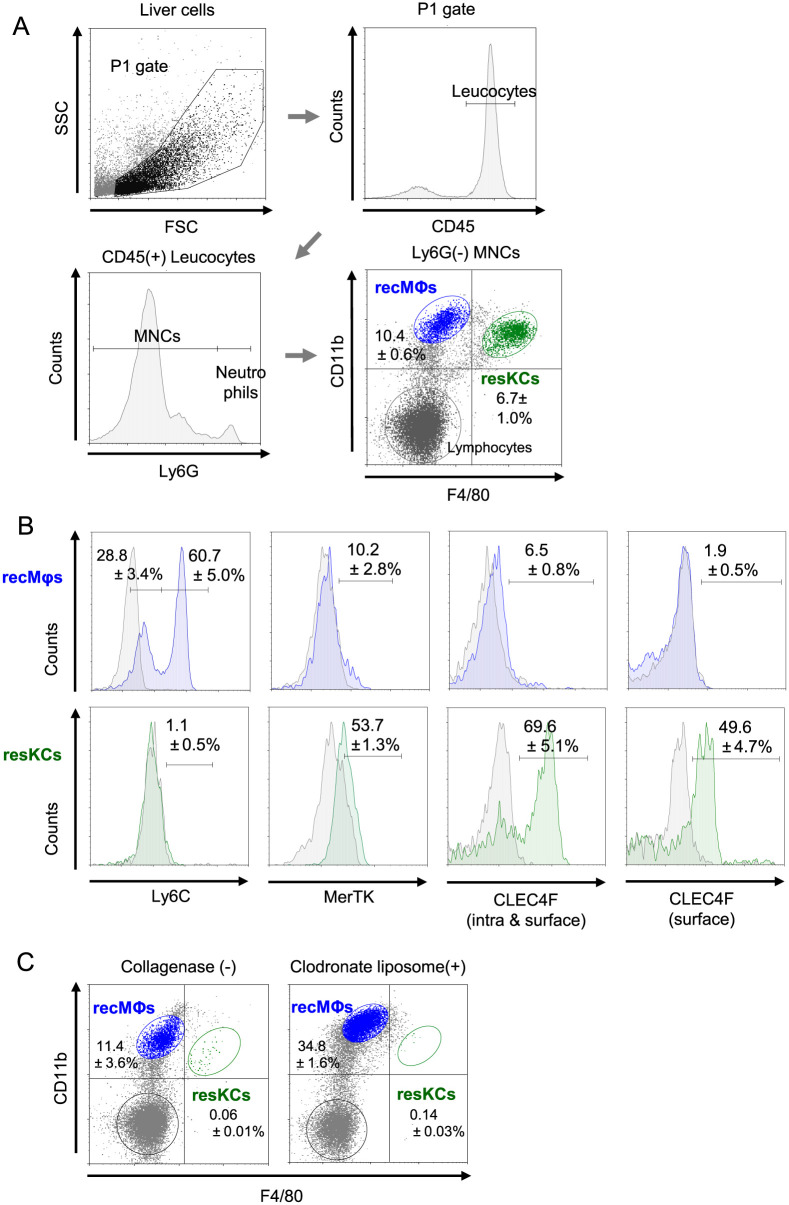
Fig 1. Flow cytometry characterization of resKCs and recMφs from mouse livers with or without collagenase pre-treatment.
(A) Gating strategy of liver recMφs and resKCs with four-color analysis. Livers from B6 mice were minced and treated with collagenase. Non-parenchymal cells were extracted and subjected to flow cytometry analysis. The extracted cells were gated with FSC and SSC, and CD45-positive immune cells were selected. Ly6G-negative cells, excluding neutrophils, were used for the experiments. Based on FACS analysis of F4/80 and CD11b expression, F4/80low CD11bhigh cells were recMφs (blue dots)., and F4/80high CD11blow cells were defined as resKCs (green dots). (n = 13). (B) The expression of Ly6C, MerTK, as well as intracellular and surface CLEC4F in recMφs (upper panels with blue histogram) and resKCs (lower panels with green histogram) was demonstrated (n = 5). (C) The FACS analysis of liver MNCs obtained without collagenase treatment (left panel) and after clodronate liposome administration (200 μl per mouse) are displayed (right panel) (n = 6). Data are presented as the mean ± SE.

