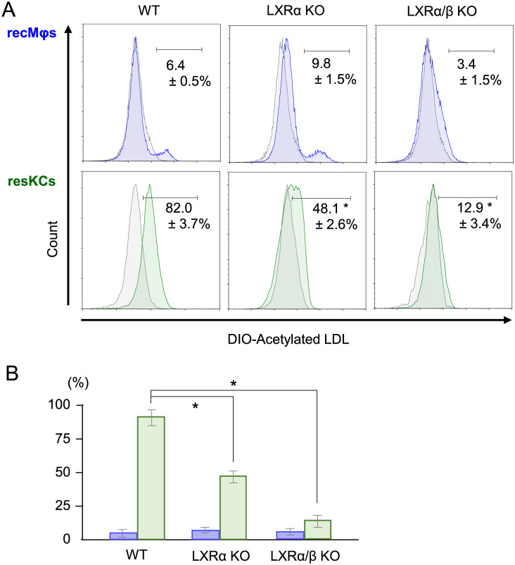
Fig 6. The decreased in vivo phagocytic activity of resKCs for acetylated LDL in LXRα KO and LXRα/β KO mice.
(A) WT mice (n = 6), LXRα KO mice (n = 3), and LXRα/β KO mice (n = 3) were injected with DIO-labeled acetylated LDL, and the endocytosis of acetylated LDL by recMφs (upper panels, blue line) and resKCs (lower panels, green line) was analyzed via flow cytometry. ResKCs and recMφs from WT mice injected with control acetylated LDL (without fluorescence) were used as negative controls (gray line). (B) Bar graph of DIO-labeled acetylated LDL endocytosis by recMφs (blue columns) and resKCs (green columns). Data are presented as the mean ± SE (*p <0.05, one-way ANOVA followed by Tukey’s multiple comparisons test).

