Figure 2.
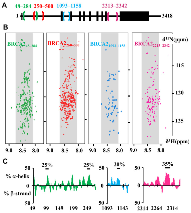
NMR analysis demonstrates that the conserved motifs BRCA248–284, BRCA2250–500, BRCA21093–1158, BRCA22213–2342 are disordered in solution. (A) Localization of the four fragments in the BRCA2 sequence. (B) 2D NMR 1H-15N Heteronuclear Single Quantum Coherence (HSQC) spectra of the four fragments. These spectra were recorded on 15N-labeled BRCA248–284 at 200 μM in HEPES 50 mM, NaCl 75 mM, EDTA 1 mM, DTT 5 mM, pH 7.0 (green, [53]), 15N-labeled BRCA2250–500 at 200 μM in phosphate 20 mM, NaCl 250 mM, pH 7.0 (red, 600 MHz CEA Saclay), 15N-labeled BRCA21093–1158 at 200 μM in 50 mM HEPES, 50 mM NaCl, 1 mM EDTA pH 7.0 (blue, 700 MHz CEA Saclay) and 15N-labeled BRCA22213–2342 500 µM, in HEPES 50 mM, NaCl 50 mM, EDTA 1 mM, pH 7.0 (pink, [54]). Cysteines of BRCA248–284 (C132A, C138A, C148A, C161A) and BRCA2250–500 (C279A, C311S, C315A, C341A, C393S, C419S, C480S) were mutated in order to favor their solubility. Grey backgrounds define the 1H frequencies typical for disordered residues. Localization of most 1H-15N peaks in the grey regions highlights the disorder propensity of these BRCA2 fragments; (C) Secondary structure propensity of three of these fragments. Propensities to form α-helices or β-strands were calculated for BRCA248–284 (green), BRCA21093–1158 (blue) and BRCA22213–2342 (pink) from their 1HN, 15NH, 13CO, 13Cα and 13Cβ NMR resonance frequencies using the ncSPC server [56]. Bold black lines underline the presence of transient secondary structure with the indicated percentage. BRCA248–284 contains two shorts transient α-helices formed by residues 100 to 110 and 255 to 260, BRCA21093–1158 exhibits one transient α-helix between residues 1122 and 1128 and BRCA22213–2342 shows one transient α-helix between residues 2292 and 2303.

