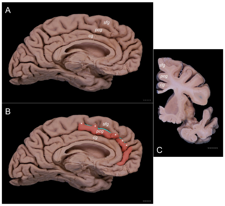
Figure 7.
An ambiguous case. Specimens are of the left cerebral hemisphere. The posterior cingulate gyrus should be classified as present based on the length of the PCS < 40 mm. However, the gyrus crosses the level of the VAC line posteriorly. (A) A medial view of this specimen. (B) A medial view of the same specimen with the PCG colored orange. The VAC and AC-PC lines are marked. The interrupted PCS is marked with teal color. (C) Coronal section of this specimen at the level of the VAC line. A separate cingulate, paracingulate, and superior frontal gyri are marked. cg—cingulate gyrus; pcg—paracingulate gyrus; sfg—superior frontal gyrus. Scale bar corresponds to 10 mm.

