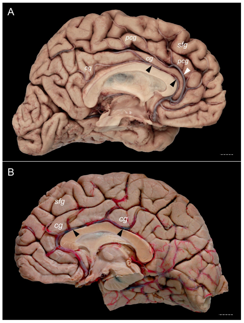
Figure 8.
The relationships of the callosomarginal and pericallosal arteries. (B) Specimen showing a prominent type of the PCG. A pericallosal artery (marked by black arrowheads) and well developed callosomarginal artery (marked by white arrowhead) are present. Pericallosal artery is thinner than the callosomarginal artery on this specimen. (A) Specimen with absent PCG. Cingulate gyrus is well developed. Pericallosal artery (marked by black arrowheads) runs within the pericallosal sulcus, over the genu and superior surface of the body of the corpus callosum. cg—cingulate gyrus; pcg—paracingulate gyrus; sfg—superior frontal gyrus. Scale bar corresponds to 10 mm.

