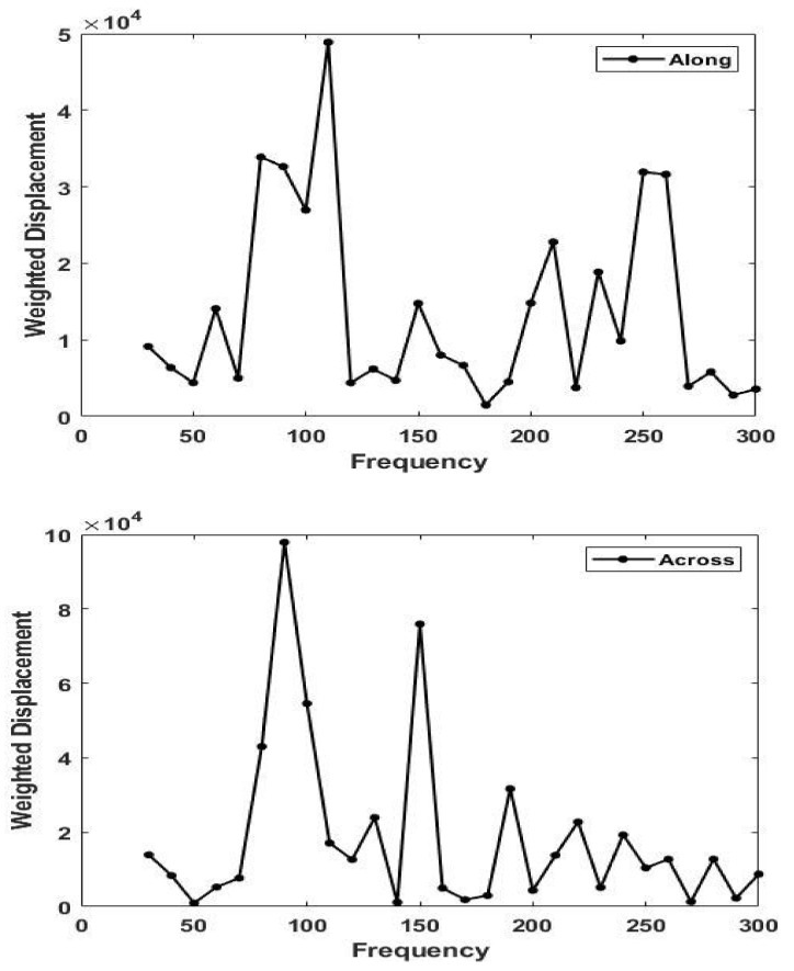
Figure 5.
Plots of weighted displacement in μm versus frequency in Hz parallel (0°) to the orientation of the collagen fibers (top) and perpendicular (90°) to the orientation of the collagen fibers (bottom) in skin in vivo at 5% strain. Note the shift in the dermal collagen peak from about 90 Hz (perpendicular) to 110 Hz (parallel). The modulus measured parallel to the collagen fibers is higher than that perpendicular to the fibers (see Equation (1)).

