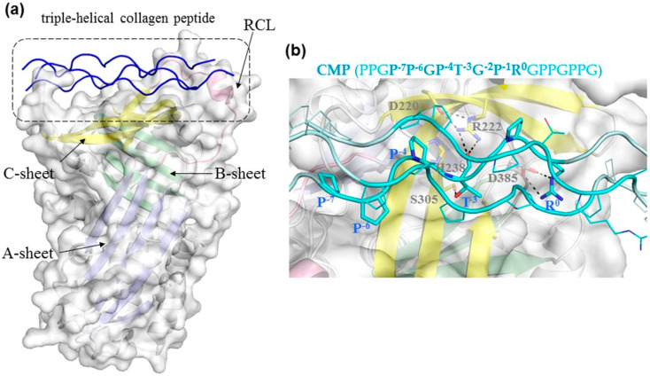
Figure 2.
A model of human HSP47 complexed with triple-helical collagen peptide. (a) The model was derived from the crystal structure of canine HSP47 complexed with collagen model peptide (CMP; PDB: 3ZHA [24]). HSP47 is shown in cartoon with three beta-sheets (A, B, C) colored in pale blue, green, and yellow, respectively, while its reactive center loop is in pink. The surface of HSP47 is shown in pale grey. The triple-helical CMP is shown in blue. (b) Close-up of the interaction between human HSP47 and the collagen peptide (PPGP−7P−6GP−4T−3GP−1R0GPPGPPG, cyan cartoon and lines). Residues of both HSP47 and CMP that are involved in interactions are shown as sticks.

