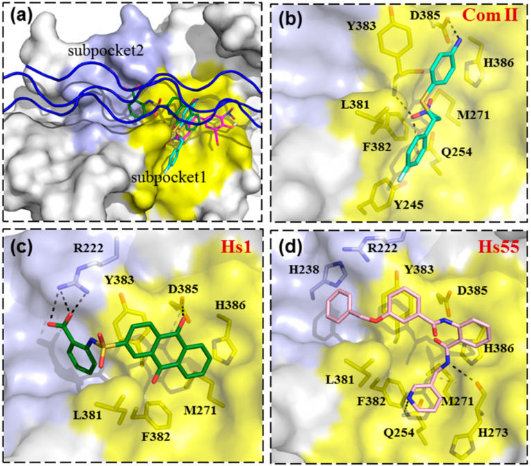
Figure 5.
Predicted binding mode of HSP47 inhibitors. (a) HSP47 is shown as surface presentations, while the collagen mode peptide (blue) is shown as a cartoon. The Arg0 binding area on HSP47 is colored yellow (subpocket 1). while the binding area for collagen residues at positions -4 and -3 is colored slate (subpocket 2). The potential binding interactions between HSP47 and the compounds Col003 (a) COM II (b), Hs1 (c), and Hs55 (d) are shown in the compounds as sticks and labeled. Compounds Col003, Com II, Hs1, and Hs55 are shown as magenta, cyan, green, and pink sticks, respectively. The black dashed lines represent the hydrogen bonds.

