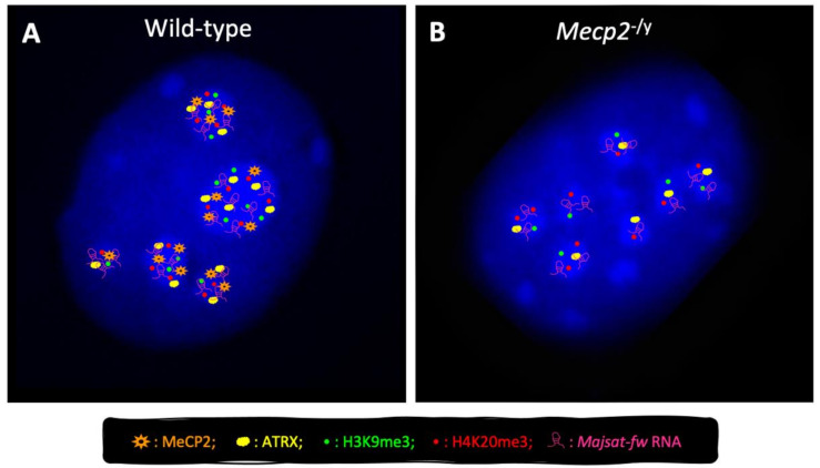
Figure 3.
Schematic representation of pericentric heterochromatin organization in nuclei of wild-type and Mecp2−/y neurons derived from differentiation of murine embryonic stem cells. Representative images of interphase wild-type (WT) (A) and Mecp2−/y (B) nuclei were depicted. Chromocenters are highlighted by intense 4′,6-diamidino-2-phenylindole (DAPI) staining (blue). In terminally differentiated Mecp2−/y cells (B), the number of chromocenters is higher and their size is lower in comparison with WT cells (A), due to the role of MeCP2 in the pericentric heterochromatin condensation (chromocenter clustering) during neural differentiation [60]. Moreover, MeCP2 contributes to the targeting of ATRX [59] and MajSat-fw transcript to chromocenters and preserves the correct deposition of H3K9me3 and H4K20me3 histone marks to pericentric heterochromatin [56].

