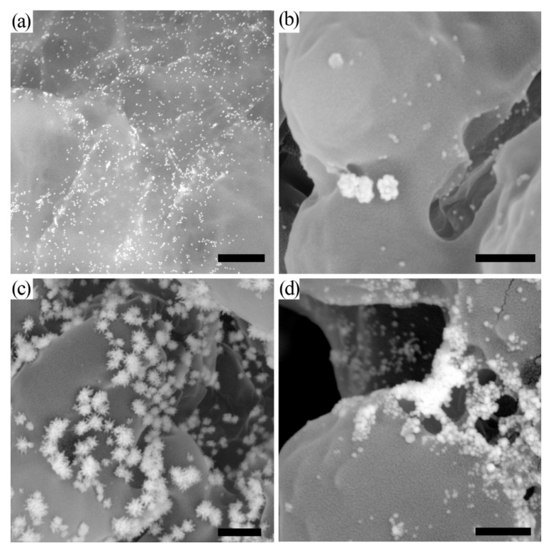
Figure 1.
Microphotographs of nanoparticles in nitrocellulose membranes. (a) Au NPs; (b) Au@Ag NPs after silver enhancement; (c) Au@Ag-Au NPs after galvanic-assisted Au deposition; (d) Au NPs after gold enhancement. The bare scale is equal to 500 nm. Microphotographs were obtained using SEM operating in back-scattered electron detection mode.

