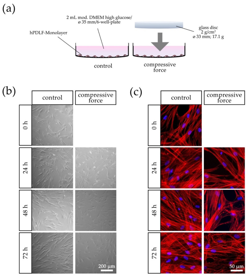
Figure 1.
Effects of compressive force (CF) on hPDLFs in terms of confluence and morphology compared to the control group at 0–72 h. (a) Schematic representation of the CF in vitro model with a sterile glass cylinder placed on the monolayer. (b) Light microscopic and (c) fluorescence microscopic phalloidin/DAPI photographs. In addition to the confluence reduction at each time point, morphological changes were especially evident after 72 h in the CF group due to the disorganization of actin filaments and punctuate accumulation of aggregates.

