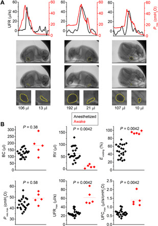
Fig. 6. Videocystometry in awake, nonrestrained mice.

(A) UFR and Pves traces with corresponding bladder images right before and after voiding in an awake, nonrestrained mouse, enclosed in a small box to ensure that the animal remains within the field of view. Indicated volume refers to Vves,spheroid, where the bladder is approximated as a spheroid (see Materials and Methods). (B) Comparison of videocystometry parameters between anesthetized (n = 22) and awake (n = 5) animals. Despite the relatively low number of awake animals in the proof-of-principle experiment, our findings indicate a significantly higher Evoiding associated with a higher maximal flow rate. Data in (B) were compared using a Mann-Whitney U test. See also movie S4.
