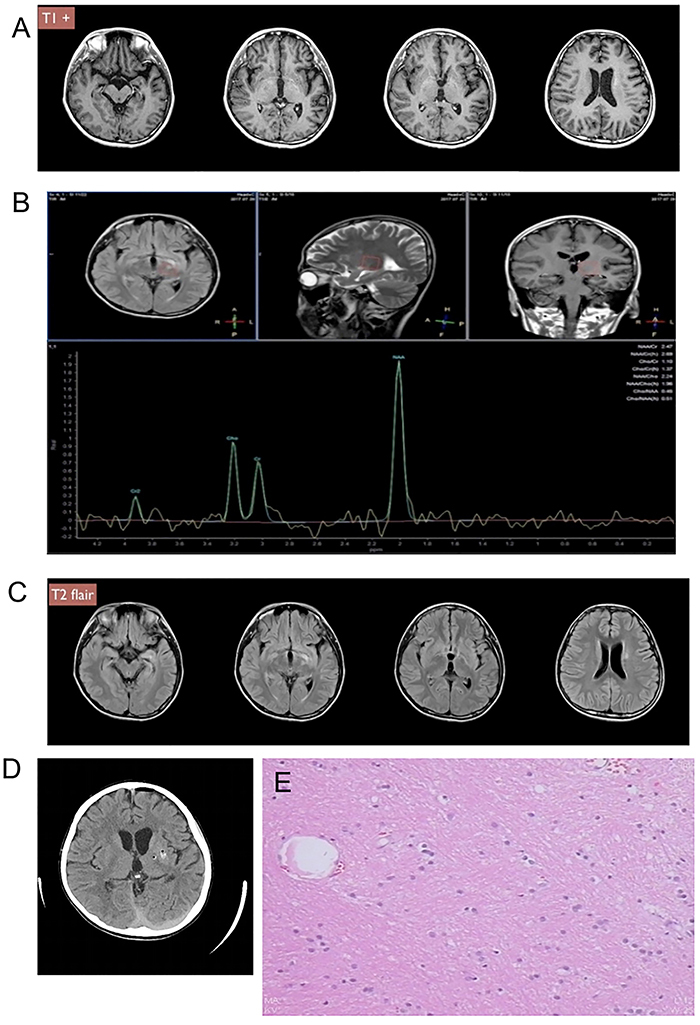
Figure 1. Case 1. A, Preoperative head T1-weighted contrast-enhanced magnetic resonance imaging (MRI) revealed left ventricle dilatation, enlarged sacral angle, and suspicious high signal on the left side of the ventricle, left basal ganglion. B, Magnetic resonance spectroscopy shows left basal ganglion NAA/Ch: 2.24, NAA/Cr: 2.69, Ch/Cr: 1.10. C, Preoperative head MRI T2 flair showed small abnormalities in the left ventricle, left midbrain, and thalamus. D, Computed tomography scan 2 h after surgery showed left basal ganglion micro-bleeding grade I and small areas of pneumocephalus. E, The pathological finding showed a single cell of a germ cell tumor.