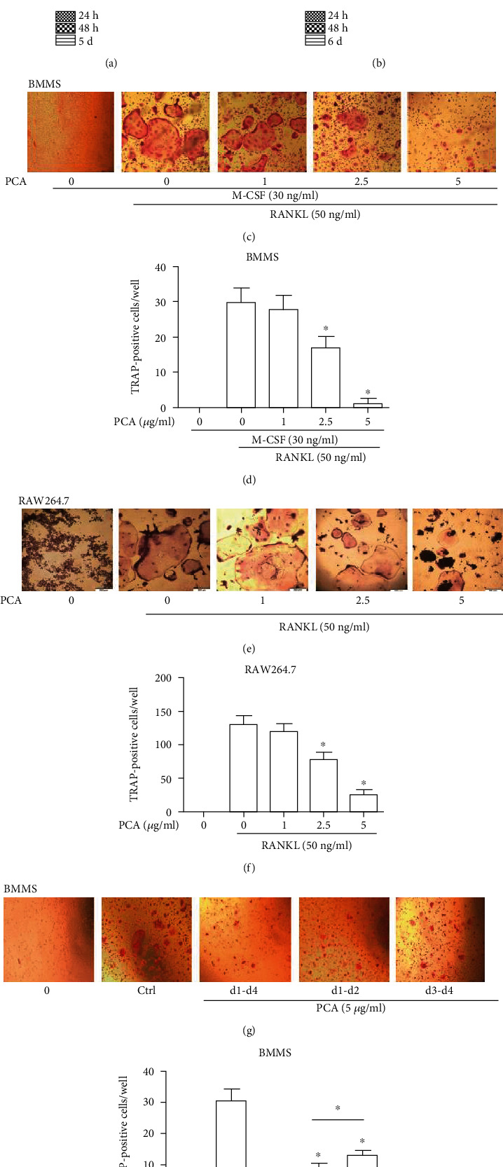
Figure 1.

PCA attenuates osteoclast differentiation induced by RANKL. (a) Viability of RAW264.7 cells exposed to PCA at different time points. (b) Viability of BMM cells exposed to PCA. (c) BMMs were induced to osteoclasts in 4 days. (d) Number of osteoclasts per well. (e) RAW264.7 cells were induced to differentiate into osteoclasts after 5 days. (f) Number of osteoclasts per well. (g) PCA (5 μg/ml) was added to BMMs at three periods during the differentiation of osteoclasts. (h) Number of osteoclasts per well. A light microscope was used to acquire photomicrographs (magnification 10 × 10). Cells that were TRAP-positive and had more than three nuclei were considered osteoclasts. The quantities are presented as the mean ± SEM values (n = 3). ∗P < 0.01 and #P < 0.05 compared with the control group.
