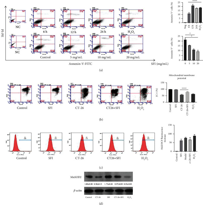
Figure 2.

SFI inhibits mitochondrial dysfunction via the restoration of mitochondrial biogenesis. (a) Representative apoptosis analysis of the different time points and SFI dosages, C2C12 mouse myoblasts cells were induced by CT-26 medium for 6, 12, and 24 h and the H2O2 for 2 h. After the CT-26 medium was stimulated for 12 h, the SFI was administered using three different dosages, 5, 10, and 20 mg/mL (∗P < 0.05 vs. control, ∗∗∗∗P < 0.0001 vs. NC values represented as the mean ± SD, n = 3). (b) SFI significantly enhanced the JC-1 mitochondrial membrane (∗∗∗∗P < 0.0001 vs. CT-26 medium; values represented as the mean ± SD, n = 3) and (c) accentuated the intracellular ROS after 6 h of incubation. (d) The expression of Mn-SOD2 using western blotting analysis after the indicated treatment. The protein densitometry data were normalized with β-actin.
