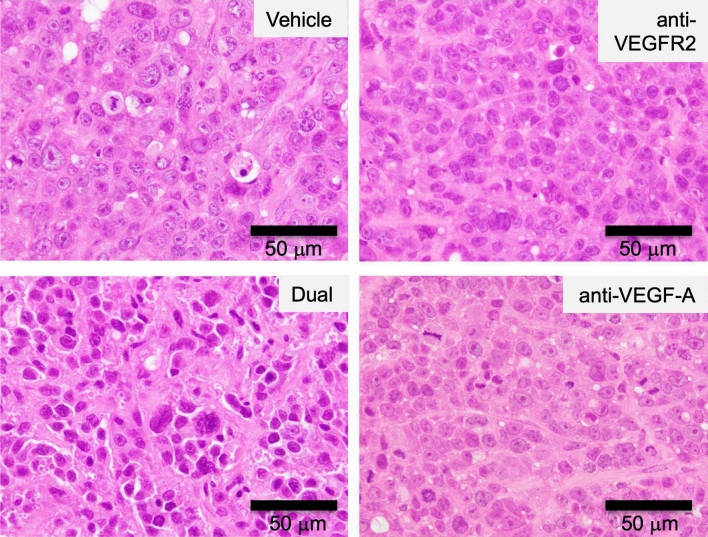
Figure 3.
Histological analysis of xenograft tumors after dual treatment with anti-VEGFR2 and anti-VEGF-A antibodies. After each treatment, xenograft tumor tissues were fixed, embedded in paraffin, and stained with hematoxylin and eosin (H&E) as described in Materials and Methods. Typical staining results in each treatment group were shown. Original magnification, ×40. The figures were generated by Microsoft Powerpoint (16.16.27) (https://www.microsoft.com/ja-jp/microsoft-365/powerpoint).

