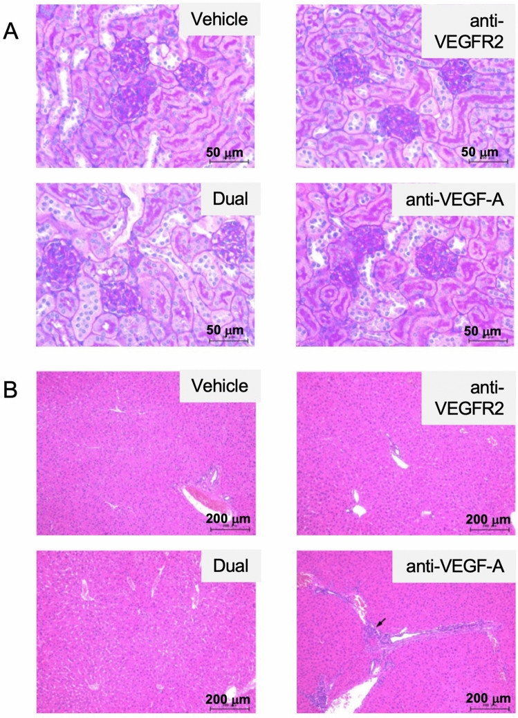
Figure 4.
Histological analysis of organs after dual treatment with anti-VEGFR2 and anti-VEGF-A antibodies in the xenograft model. (A) Effect of treatment with anti-VEGFR2 and anti-VEGF-A antibodies on mouse kidneys. After each treatment, kidney tissues were fixed, embedded in paraffin, and stained with Periodic acid–Schiff (PAS) as described in Materials and Methods. Typical staining results in each treatment group were shown. Original magnification, ×40. (B) Effect of treatment with anti-VEGFR2 and anti-VEGF-A antibodies on mouse livers. Liver tissues were fixed, embedded in paraffin, and stained with hematoxylin and eosin (H&E) as described in Materials and Methods. Typical staining results in each treatment group were shown. The arrow indicates slight bile duct hyperplasia. Original magnification, ×10. The figures were generated by Microsoft Powerpoint (16.16.27) (https://www.microsoft.com/ja-jp/microsoft-365/powerpoint).

