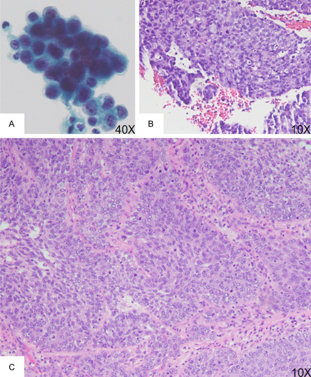
Figure 2.

Urine cytology, ureteroscopic biopsy and nephroureterectomy from a representative case of HG UTUC in the renal pelvis. A. Urine cytology specimen shows tumor cells with enlarged nuclei, nuclear hyperchromasia, irregular nuclear membrane and increased nuclear-to-cytoplasmic ratio (> 0.7). B. Ureteroscopic biopsy shows papillary architecture with loss of cellular polarity, enlarged nuclei with nuclear hyperchromasia, and increased mitotic activity and apoptotic bodies. C. Section of tumor from a nephroureterectomy specimen shows invasive tumor nests with disorganized tumor cell arrangement with enlarged nuclei, prominent nucleoli, and increased mitotic activity.
