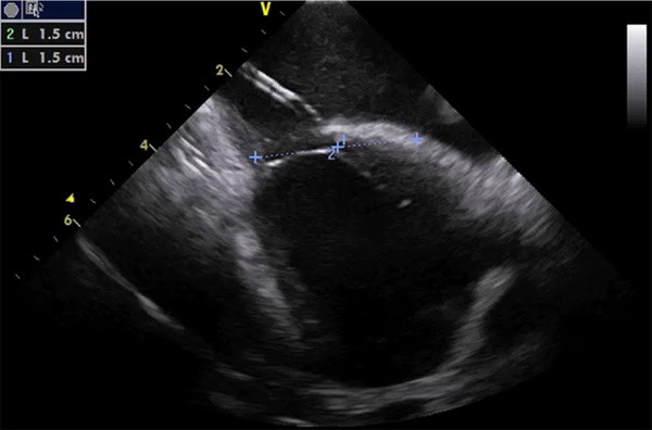
Figure 2.

ICE imaging for guidance of PFO closure, showing the delivery catheter through a tunneled defect, positioned from right (above) to left (below) atrium. As showed, a proper sizing can be performed with two measurements drawn from the center (1 and 2), simulating the final position of the device (30 mm in this case).
