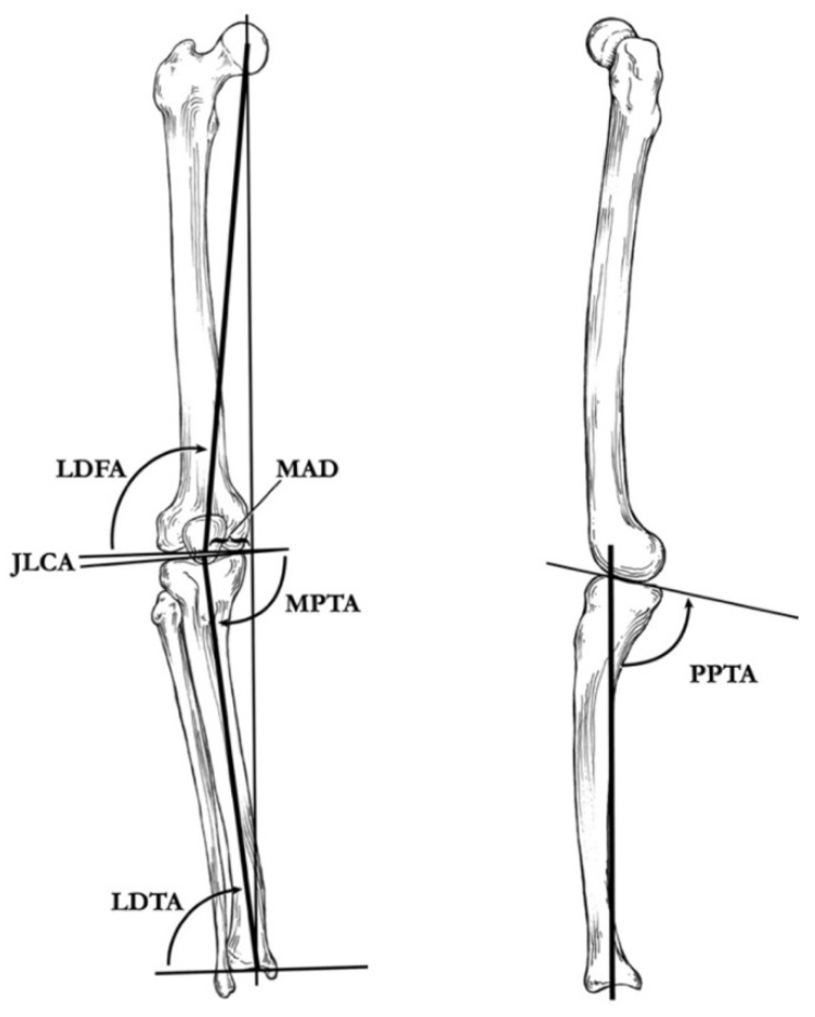
Figure 6.
(Left): Coronal plane illustration of the lower extremity, demonstrating the mechanical axis deviation (MAD), the lateral distal femoral angle (LDFA), the joint–line congruency angle (JLCA), the medial proximal tibial angle (MPTA), and the lateral distal tibial angle (LDTA). (Right): Sagittal plane illustration of the proximal part of the tibia and the distal part of the femur, demonstrating the posterior proximal tibial angle (PPTA) [29].

