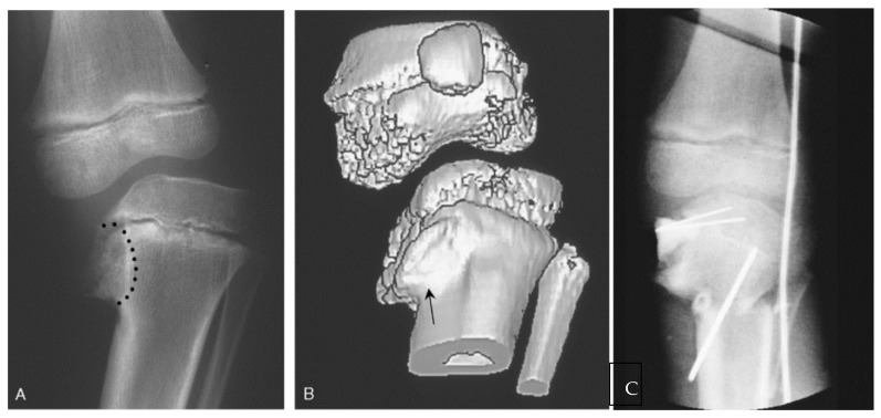
Figure 13.
(A) A 6-year-old female with untreated stage IV ITV; the metaphyseal bone and Blount lesion to be excised is outlined; (B) 3D CT showing extent of abnormal depressed epiphyseal bone anteriorly. The "cleft" on the anterior surface of the metaphysis (arrow) is where the initial resection for epiphysiolysis begins. (C) After interposition of cranioplast at the site of resection and stabilization of a closing wedge tibial osteotomy. The bovie cord defines the lateral mechanical axis [65].

