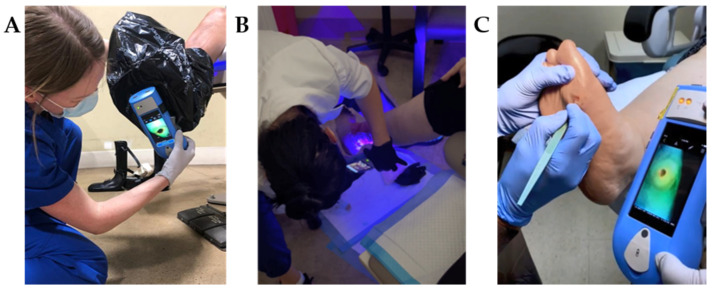
Figure 1.
Fluorescence imaging procedure for wound bacterial presence, location and load performed across multiple sites of service. (A) When in a bright environment, a DarkDrape® is attached and used to create adequate darkness. The drape is positioned around the anatomy and the fluorescence imaging device is positioned at an appropriate distance (8–12 cm) from the wound prior to focusing and capturing an image. (B) The DarkDrape® is not required in a darkened environment. For large wounds wrapping around the anatomy, as shown here, multiple fields of view will be imaged and interpreted. (C) Fluorescence images can be immediately interpreted at the point-of-care to inform treatment. Multiple images may be acquired per visit to assess efficacy of treatments performed. This panel shows the image informing a physician’s debridement.

