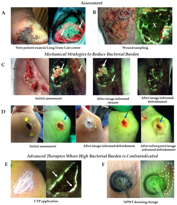
Figure 3.
Use of fluorescence imaging across multiple sites of service. (A) Fluorescence images captured at initial assessment of a patient in long term care with a diabetic foot ulcer revealed significant bacterial burden around the wound (white arrows) that changed the plan of treatment put in place for this patient. (B) Images of a venous leg ulcer prompted clinician to collect wound biopsy from region of cyan fluorescence (denoted by red dotted circle), rather than the wound center (denoted by an ‘X’); sample was positive for Pseudomonas aeruginosa. (C) Patient with an incisional hip wound following surgery had red fluorescence (white arrows) from bacteria in and around wound. The wound was washed vigorously (HOCL cleanser) and re-imaged. Persistence of red fluorescence prompted additional cleansing and debridement in regions of red fluorescence. Red fluorescence diminished after image-informed mechanical strategies. (D) Red fluorescence from bacteria was detected in a diabetic foot ulcer, highlighted by blue arrow, prompting debridement. Initial debridement revealed a larger amount of bright red fluorescence in the callus tissue surrounding the wound. Additional debridement was performed to remove bacteria laden tissue. (E) A diabetic foot ulcer that had previously received multiple CTP applications underwent fluorescence imaging prior to application of another CTP. Images revealed the presence of cyan fluorescence (white arrows) indicative of Pseudomonas aeruginosa around the wound edge; CTP application was withheld until cyan signal was eliminated. (F) NPWT was applied to an appendectomy abscess. Fluorescence images were captured during a dressing change and indicated presence of red fluorescence around the wound (denoted by dashed white dotted line). Imaging informed mechanical bacterial removal prior to placement of a new dressing. CTP, cellular tissue product; NPWT, negative pressure wound therapy.

