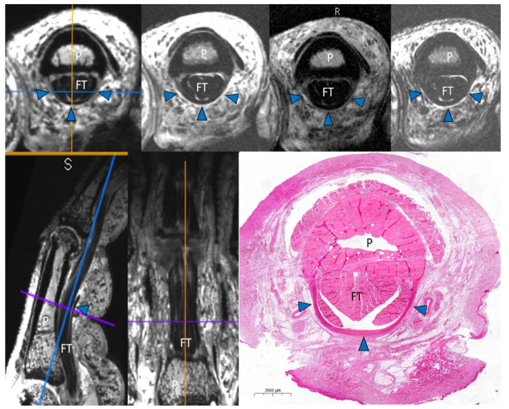
Figure 1.
Example of the study protocol with an intact A2 pulley. Three-dimensional double echo steady-state (3D DESS) axial multiplanar reconstruction (MPR), axial PD fs, T1 turbo spin echo (TSE), and T2 TSE scans (above). 3D DESS sagittal and coronal MPR. Histological correlation (below). P = phalanx. FT = flexor tendon. Arrowheads = intact A2 pulley.

