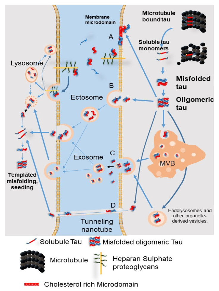
Figure 6.
The secretion pathways of pathological tau protein. (A) Direct secretion of tau through the plasma membrane involves clustering of tau at the plasma membrane, interaction of cholesterol/sphingomyelin-rich membrane microdomains with specific lipids, permeation into the plasma membrane, and release from the plasma membrane by cell surface heparin sulfate proteoglycans. (B) Tau is secreted into ectosomes, which are larger and have a different composition than the exosomes released from the plasma membrane. Both ectosomes and exosomes function in the same manner after they are released from the cell, fusing to the target cell or being endocytosed. (C) Secretion of tau by exosomes and organelle hitchhiking. Tau may be secreted when the membrane of late endosomes exits inward to form luminal vesicles of the multivesicular body (MVB), which are packed into exosomes, and when the MVB membrane fuses with the plasma membrane. Organelle hitchhiking pathways that may be involved in the secretion of tau and other misfolded cytoplasmic proteins include secretory endolysosomes associated with the autophagy–lysosome pathway. The misfolding-associated protein secretion (MAPS) pathway involves chaperone-mediated uptake of misfolded cytoplasmic proteins into the endoplasmic reticulum, followed by fusion of endolysosomal vesicles with the plasma membrane and secretion, thereby releasing vesicle-free tau into the extracellular space. (D) Tau seeds are transferred between cells via tunnel-like nanotubes that directly connect the cytoplasm of two adjacent cells. MVB: multivesicular bodies. (Revised from Brunello, C.A. et al., Cell Mol. Life Sci. 2020, 77, 1721-1744 [60]).

