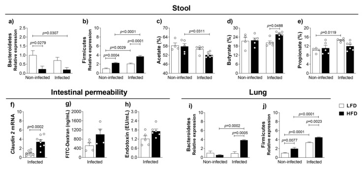
Figure 3.
Obesity-induced dysbiosis in the lung and gut. C57BL/6 mice were fed with the LFD or HFD for 8 weeks and infected with M. tuberculosis by intra-tracheal (it) route. At 4 weeks post-infection (12 weeks of feeding), mice were evaluated. (a) Bacteroidetes and (b) Firmicutes relative expression from feces. Short-chain fatty acids, (c) acetate, (d) butyrate and (e) propionate were evaluated in stool samples (n = 5–6). Intestinal permeability evaluated by (f) relative expression of claudin-2 mRNA in the ileum, (g) FITC-Dextran (ng/mL) and (h) endotoxin (EU/mL) in serum. Data are representative of two independent experiments for claudin-2 and one experiment for FITC-Dextran and endotoxin (n = 4–8/group). (i) Bacteroidetes and (j) Firmicutes relative expression from lung samples (n = 7–12). Data are representative of three independent experiments for Bacteroidetes analysis and two independent experiments for Firmicutes analysis. Data are representative of two independent experiments and are expressed as means ± SD. Bars show the significant difference between groups (p < 0.05).

