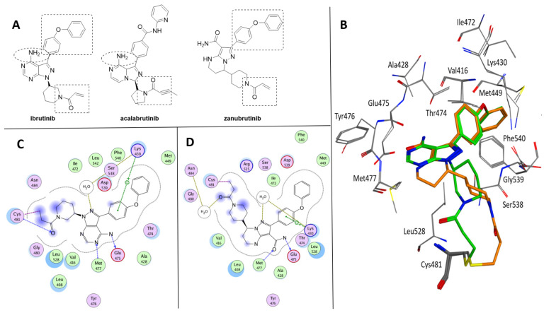
Figure 2.
(A) Chemical structure of ibrutinib, acalabrutinib and zanubrutinib. The shared subportions are evidenced. (B) Superposition of Btk crystallized with ibrutinib (green, PDB code 5P9J) and zanubrutinib (orange, PDB code 6J6M). (C) Ligplot of the Btk–ibrutinib complex. (D) Ligplot of the Btk–zanubrutinib complex.

