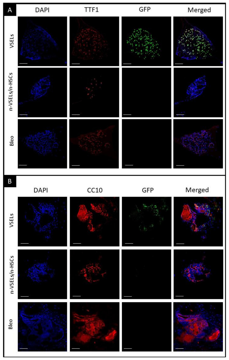
Figure 7.
Detection of airway epithelia and AT2 cells in organoids. Immunofluorescent staining of representative organoid sections from each of transplant groups as indicated. Scale bars represent 100 µm. Panel A: blue—DAPI, red—TTF1, green—GFP, and merged. Colocalization of DAPI, GFP, and TTF1 indicates donor VSEL derived AT2 cells, representing maintenance of proliferation and differentiation; Panel B: blue—DAPI, red—CC10, green—GFP, and merged colors. Colocalization of DAPI, GFP and CC10 indicates that donor VSEL-derived BASCs self-renew and form organoids, which proves their full physiological abilities. Note that CC10+ cells are GFP−, as GFP is expressed from the SPC promoter. CC10 is cytoplasmic and secretory protein what can be observed in stained Matrigel (cell-free, no DAPI nuclei) in addition to individual cells.

