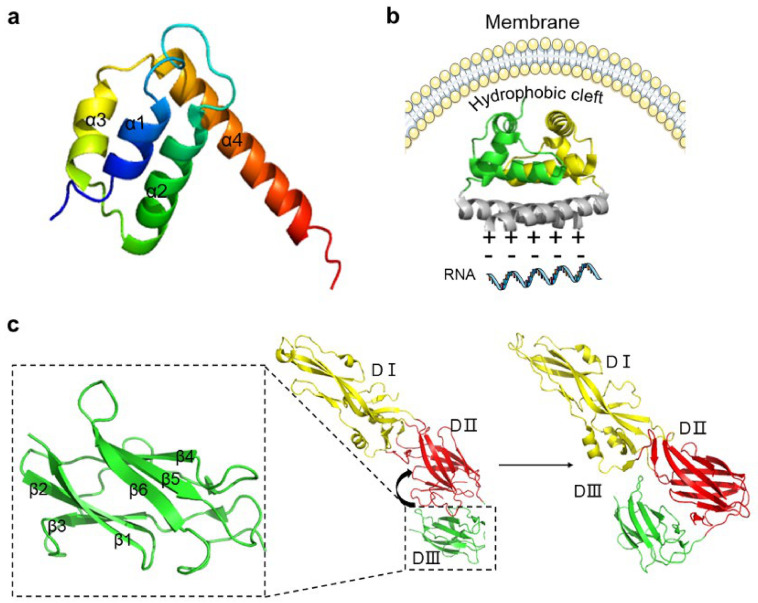
Figure 2.
The structures of capsid protein (C protein) and envelope protein (E protein). (a) The structure of the dimer of C protein of dengue virus (DENV; Protein Data Bank [PDB]: 1R6R DENV). (b) C protein tetramer. C protein is arranged in a symmetrical form of 2:2:2, forming a hydrophobic channel in the middle of the tetramer (PDB: 1SKF West Nile virus [WNV]). (c) The three domains of E protein; the detail of DIII is shown in the enlargement of the framed area. There is a flexible short chain between DII and DIII; the arrow indicates the conformational change of DIII towards DII during maturation (PDB: 1TG8 DENV).

