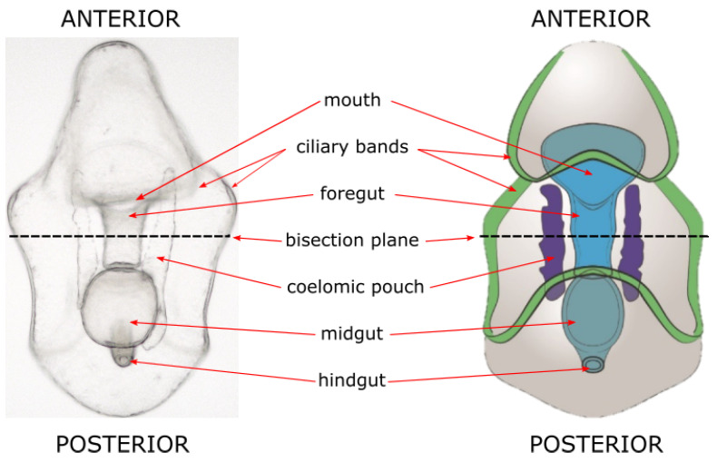
Figure 1.
Sea star bipinnaria larval morphology. Left, a light microscopy image of a larva of a sea star (Patiria miniata). Large structures are easy to identify and tissues are transparent, making both light and fluorescent microscopy advantageous. Right, a schematic of major sea star larval structures.

