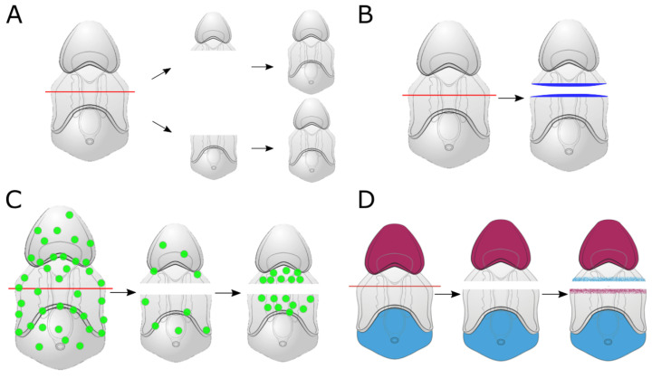
Figure 2.
(A) graphical summary of the results from Cary et al. (2019) [41] examining mechanisms of larval sea star regeneration. Sea star larvae proportion their body size following bisection. This results in regenerated larvae that have similar proportions to that of an uncut larva but a smaller overall size. (B) Soon after bisection, the larval epithelium wound closes and expression of wound-induced genes is seen at the site of damage (blue area). This includes the expression of genes such as Elk and Egr and the localization of phosphorylated-ERK. (C) Subsequent to a reduction in overall cell proliferation early in regeneration (green circles), a cluster of proliferative cells emerges specifically at the injury site, akin to a regeneration blastema. (D) Loss of expression of genes along the anterior-posterior (AP) axis is recovered as regeneration proceeds. Many of these genes are components of the canonical Wnt-signaling pathway.

