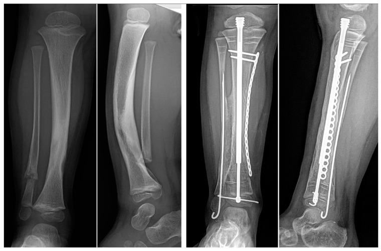
Figure 9.
AP and lateral radiographs of a patient with Paley type 2A CPT. The preoperative images on the left show an intact tibia with fibular pseudarthrosis. A tibial osteotomy was made at the apex of the deformity, straightened, and fixed with the standard cross-union protocol. The postoperative images on the right show a well-healed osteotomy and cross-union with normal tibial alignment.

