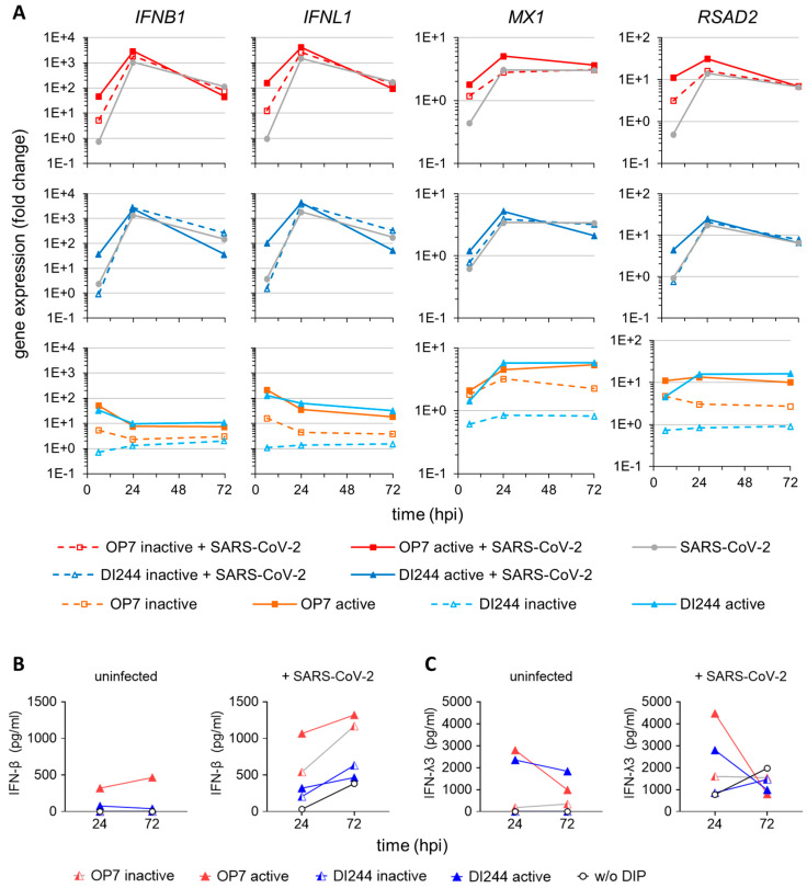
Figure 3.
Stimulation of IFN-induced antiviral activity by IAV DIP infection. SARS-CoV-2-infected Calu-3 cells (MOI = 0.03) were treated with IAV DIPs (DI244 or OP7) at 1 hpi. For DI244 and OP7 infection, 10% (v/v) (100 µL culture volume) of highly concentrated cell culture-derived DIP material was used [29,30]. At indicated times post-infection, infected cells were lysed to allow for total RNA extraction, required for (A) gene expression analysis. In addition, supernatants were sampled for (B,C) quantification of secreted IFNs. Illustration includes data from one experiment. (A) Gene expression analysis of SARS-CoV-2 and IAV DIP co-infection. Transcript levels were quantified by real-time RT-qPCR and expressed as fold change (relative to untreated, uninfected cells). MX1, MX dynamin-like GTPase 1; RSAD2, radical S-adenosyl methionine domain-containing 2. (B,C) Host cell IFN production. Protein levels of IFN-β (B) and IFN-λ3 (C) were assessed using ELISA.

