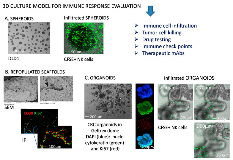
Figure 1.
Representative 3D models to study NK-tumor cell interactions. (A) Colorectal cancer (CRC) cell spheroids from DLD1 CRC cell line (left) infiltrated with NK cells (right) labelled with the green fluorescence probe carboxy fluorescein succinimidyl ester (CFSE). (B) Collagen scaffolds repopulated with mesenchymal stromal cells and Hodgkin’s lymphoma cells (scanning electron microscopy, SEM, upper) or analyzed by immunofluorescence (IF, lower) for the expression of CD30 antigen (HL cells, red) and Ki67 (proliferating antigen, green); (C) CRC organoids in a GeltrexTM dome (left), or labelled (middle) with 4′,6-diamidino-2-phenylindole (DAPI, blue), cytokeratin 2 (green), Ki67 (red), or merge (lower image); organoids analyzed by confocal microscopy upon NK cell (green labeled with CFSE) infiltration at different Z-stages (right). The dimension bars are shown in each panel.

