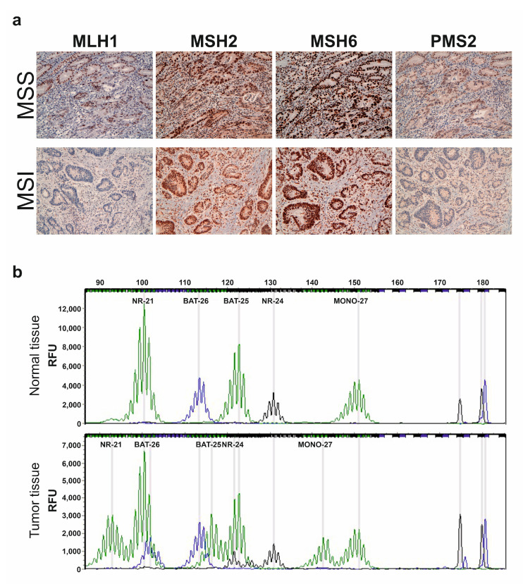
Figure 1.
Representative IHC staining for MMR proteins and electropherograms of multiplex PCR analysis using the MSI Analysis System Version 1.2 kit. (a) MLH1, MSH2, MSH6 and PSM2 expression in FFPE samples of a MSS and a MSI patient by IHC (original magnification 20×). The MSS patient shows normal expression of all MMR proteins. Deficiency in MLH1 and PSM2 proteins is observed in the MSI patient; (b) Multiplex PCR electropherograms show profiles of 5 quasimonomorphic microsatellites (NR-21, BAT-26, BAT-25, NR-24 and MONO-27) in the normal and tumor tissue of the same patient. The tumor tissue shows additional peaks that are absent in the normal tissue of all microsatellites analyzed, revealing an MSI profile.

