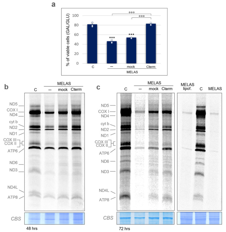
Figure 2.
Cterm rescues viability and mitochondrial translation in MELAS cybrids. (a) Viability of wild-type control cybrids (C) and MELAS cybrids either non-transfected (-) or transiently transfected with an empty pcDNA6.2 vector (mock) or a Cterm-overexpressing vector (Cterm) evaluated after 24 h-incubation in galactose medium. The number of viable cells in galactose was normalized to the number of viable cells in glucose. Results are presented as the mean ± S.D. Statistical analysis was performed on three independent biological replicates using two-tailed Student’s t test; asterisks (***, p < 0.001) indicate the significance of the decreased viability in MELAS cells with respect to the control. Normal distribution of MELAS samples was confirmed by the Shapiro–Wilk test and means were simultaneously compared by one-way ANOVA (α: 0.05) followed by post-hoc Tukey’s test; degree symbols (°°°, p < 0.001) refer to the significance of the increase in cybrids overexpressing Cterm with respect to the non-transfected and mock. Individual data points are depicted by white diamond-shaped dots. (b) Metabolic [35S]-methionine labelling of mitochondrial translation products performed in wild-type cybrids (C) and in MELAS cybrids detailed in (a). Pulse-labelling (1 h) was carried out 48 h after transfection; total cell protein (20 µg) were separated by 15% SDS-PAA gels. Mitochondrially encoded polypeptides were assigned, as in [24]. Coomassie blue staining (CBS) of the gel was used as loading control. (c) Metabolic [35S]-methionine labelling as in (b), except that labelling was performed 72 h after transfection (left panel). Labelling of MELAS cells treated with the transfection reagent only (lipof.) was also carried out (right panel).

