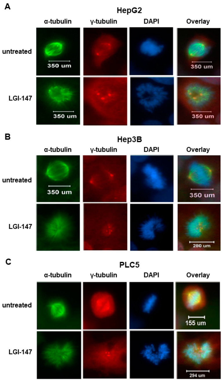
Figure 3.
(A–C) Abnormal monopolar spindle formation in HCC cells under LGI-147 treatment during prometaphase. After 24 h treatment with the vehicle or 50 pM of LGI-147, the HCC cells were fixed and stained with anti-α-tubulin (green) and anti-γ-tubulin antibodies (red) and DAPI (blue). The images were captured using a confocal microscope (63× objective).

