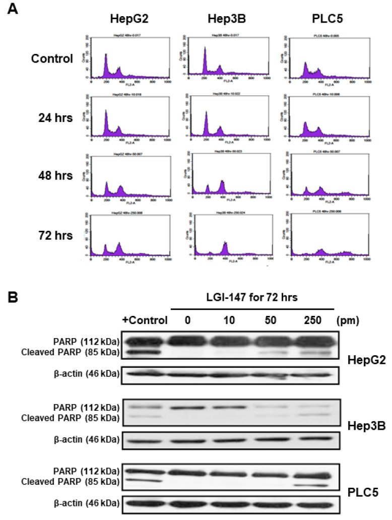
Figure 4.
(A) Cell cycle disturbance in the HCC cells. Cells were treated with 50 pM LGI-147 for 24 to 72 h, and then stained with propidium iodide. DNA content was analyzed using the flow cytometry. Data shown are representative of three independent experiments. (B) Cell apoptosis. The HCC cells were treated with a vehicle, positive control (1 μM doxorubicin), or indicated concentrations of LGI-147 for 72 h. PARP cleavage was detected using Western blotting.

