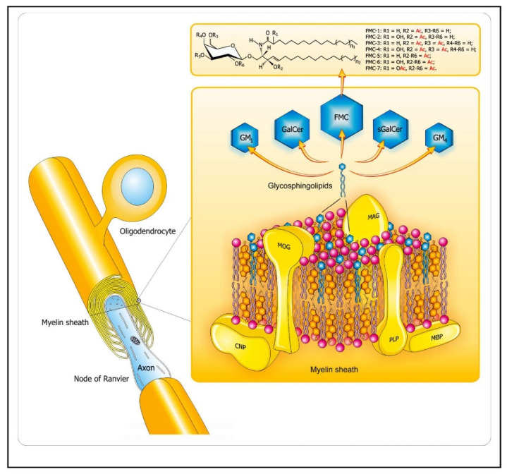
Figure 2.
Lipid distribution in the CNS myelin sheaths. The diagram depicts arrangement of complex lipids (cholesterol, phospholipids, and glycosphingolipids) within the most abundant proteins (PLP, MBP) in CNS myelin. The relative molar constancy of lipids: cholesterol/phospholipids (PLs)/galactosylceramide (GalCer) is 2:2:1. Proteins are marked in yellow, and the comprising lipids are as follows: cholesterol in orange, PLs in pink, and the glycosphingolipids (FMC, fast migrating cerebrosides; GalCer, galactosylceramide; GM1, mono-sialoganglioside; GM4, sialosyl-galactosylceramide; sGalCer, sulfatide) in blue. Structures of unique sphingosine 3-O-acetylated-GalCer glycolipid series, namely, acetyl-cerebrosides (FMCs) are shown at the top. Abbreviations: CNP—2′3′-cyclic-nucleotide 3′-phospodiesterase, MAG—myelin-associated glycoprotein, MBP—myelin basic protein, MOG—myelin oligodendrocyte glycoprotein, PLP—proteolipid protein. Adapted from [5].

