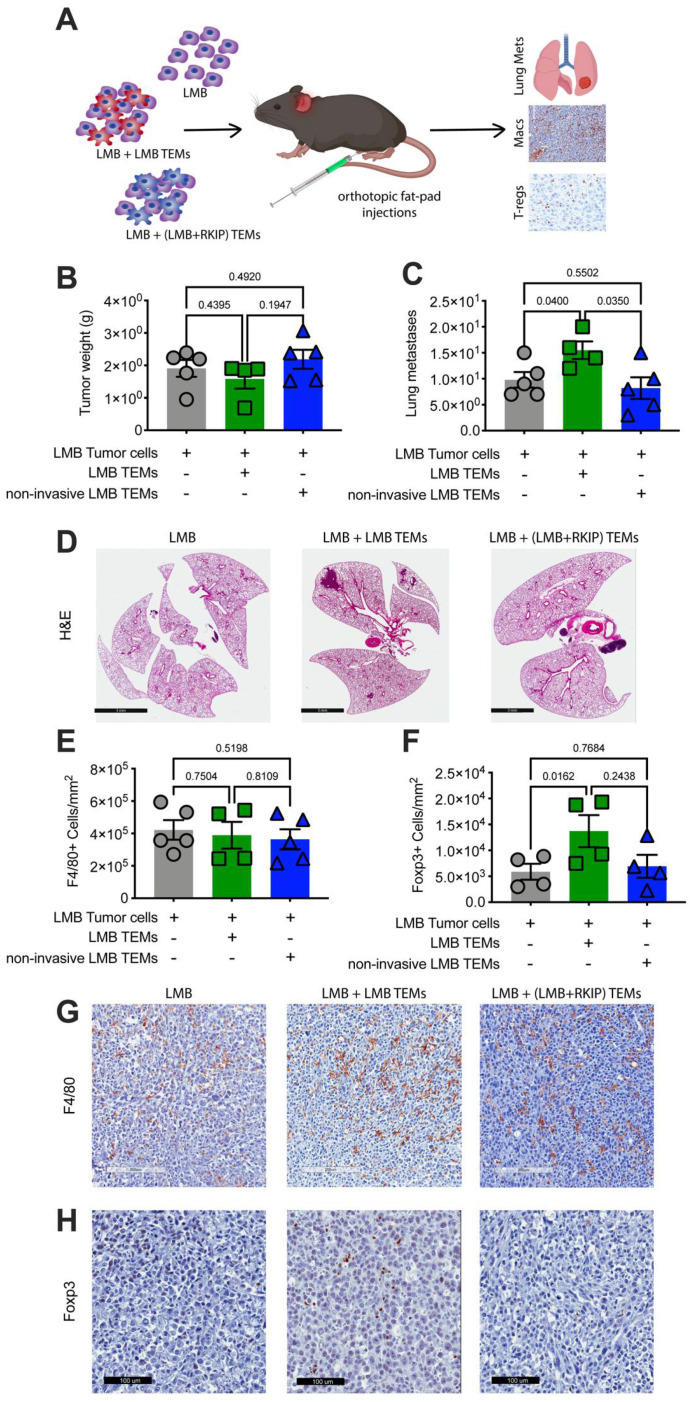
Figure 6.
Tumor EVs promote metastasis through macrophages. (A) Either 0.5 × 10⁶ LMB EV programmed or LMB (overexpressing RKIP) EV programmed TEMs were co-injected with tumor cells as indicated. Final tumor weights (B) and number of metastases (C) per mouse are shown for LMB tumors alone (grey, N = 5), LMB tumor cells with LMB programmed TEMs (green, N = 4), and LMB tumor cells with LMB (RKIP overexpressing, non-metastatic) programmed TEMs (blue, N = 5). (D) Representative images of lung metastases from each group. Final number of macrophages (E) and Foxp3+ T-regs (F) per mouse are shown for LMB tumors alone (grey, N = 5), LMB tumor cells with LMB programmed TEMs (green, N = 4), and LMB tumor cells with LMB (RKIP overexpressing, non-metastatic) programmed TEMs (blue, N =5). Representative images of F4/80+ macrophages (G) and Foxp3+ T-regs (H) from each group. (All p-values were calculated utilizing a t-test).

