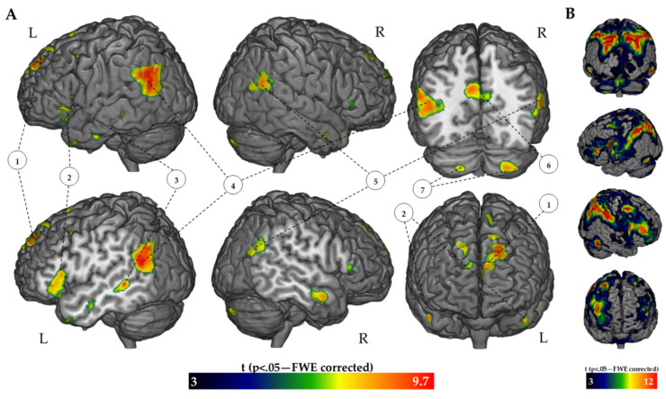
Figure 1.
Threshold activation maps of the FR > BC contrast (A) and of the BC > FR contrast (B). The regions are displayed on the 3D rendered MNI reference brain (statistical threshold of p < 0.05 FWE-corrected; cluster size: >50). 1: Bilateral Superior/Medial Frontal Gyrus; 2: Left Inferior Frontal Gyrus; 3: Left Middle Temporal Gyrus 4: Left IPL; 5: Right IPL; 6: PCC and Precuneus; 7: Bilateral Cerebellum.

