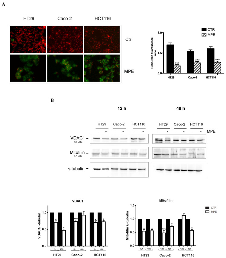
Figure 3.
MPE treatment causes dissipation of mitochondrial membrane potential (Δψm) and changes in mitochondria-associated proteins. (A) JC-1 staining on colon cancer cells untreated and treated with 360 μg/mL MPE for 48 h. Red fluorescence, suggestive of a high mitochondrial membrane potential (preserved Δψm favors JC-1 aggregates), was observed in almost all untreated cells, whereas green fluorescent signals, indicative of low mitochondrial membrane potential (depolarization favors JC1 monomers), occurred in MPE-treated colon cancer cells. Merged images were obtained as reported in the Materials and Methods section, and taken at 200x magnification (original). Quantification of green and red fluorescent cells (expressed in percentages) is reported in the bar chart. Data were from three independent experiments, each with 50 counted cells. The data are reported as mean ± SD. (***) p < 0.001 versus untreated control. (B) Western blot analysis of the mitochondria-localized proteins. Cells were treated for 12 h and 48 h, then total cell extracts were subjected to Western blot analysis and tested for VDAC1 and mitofilin. The correct loading was checked for γ-tubulin protein. The densitometric analysis data reported are the mean of results obtained via three separate experiments that were normalized to the γ-tubulin protein. (*) p < 0.05 and (***) p < 0.001 versus untreated control.

