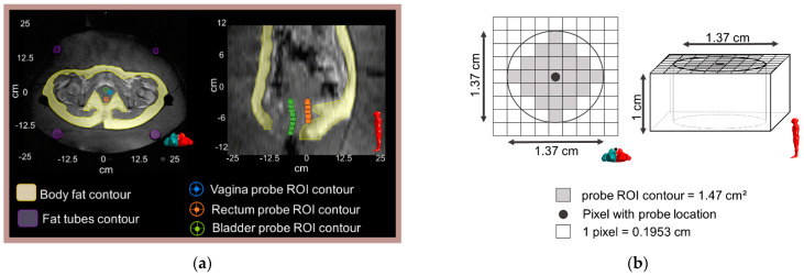
Figure 2.
(a) An axial and a sagittal T1-weighted MR image of the pelvic are shown along with an illustration of the location of the Bowman probes points and the corresponding delineated ROIs: vagina, rectum, and bladder. The left and right images correspond to the middle axial slice (z = −2 cm) and middle sagittal slice (x = 0 cm), respectively. Body fat and fat-like tube ROIs are indicated in the axial MR image. (b) Schematic representation of the probe ROI contour in the axial and sagittal view.

