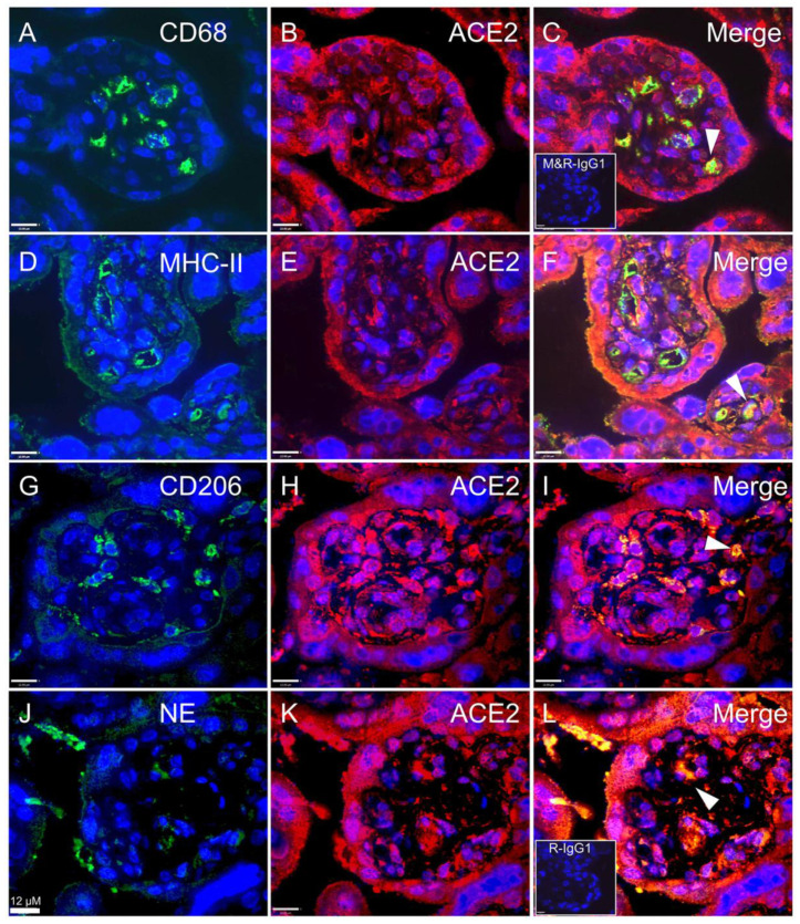
Figure 6.
Co-localization of SARS-CoV-2 associated cell-entry protein, ACE2, within specific immune cell populations in ChA placenta. Representative images of ACE2, CD68, M1, M2 and NE staining in immunofluorescence (ACE2; red color and M1, M2, macrophage and NE; green color), macrophage co-staining with ACE2 (A–C) M1 co-staining with ACE2 (D–F), M2 co-staining with ACE2 (G–I) and neutrophil co-staining with ACE2 (J–L). Co-localization of ACE2 and markers of macrophage and neutrophil confirmed the ACE2 localization within placental macrophages and neutrophils. ACE2 expressing immune cells were present in the villous stroma and adjacent to fetal blood vessels. Only a subset of each of immune cells expressed ACE2. Arrows show ACE2 staining within CD68, M1, M2 and NE stained cells. Sections were counter-stained with DAPI (blue color) or co-staining (yellow color). Inserts (bottom left) in (C) mouse and rabbit IgG1 isotype control and (L) rabbit IgG1 isotype control staining. n = 3/group. Scale bar represents 12 μm.

