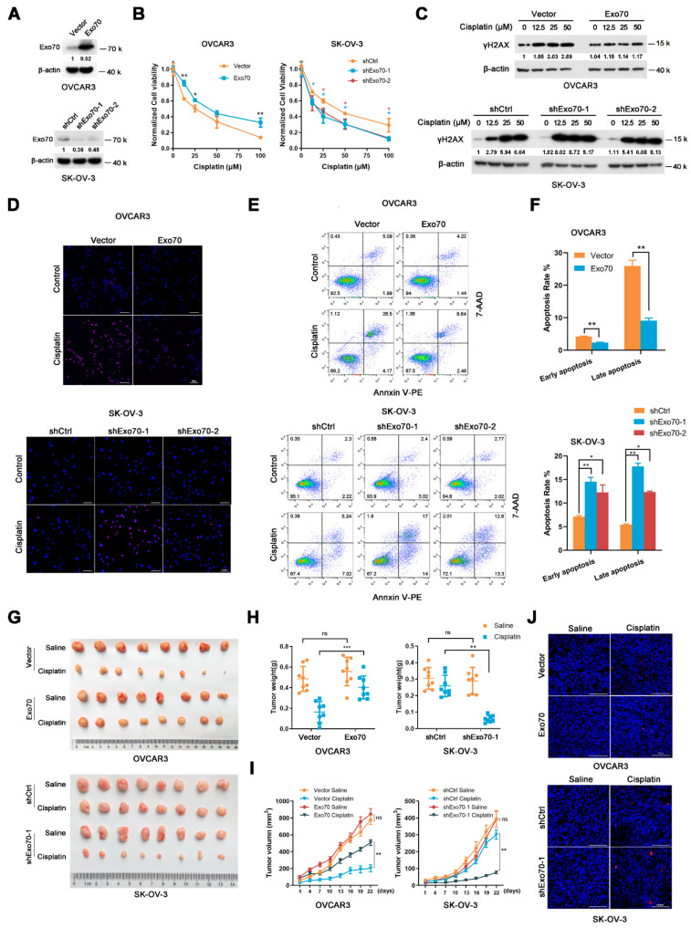
Figure 2.
Exo70 affects cisplatin sensitivity of EOC in vitro and in vivo. (A) Western blotting showed the overexpression of Exo70 in OVCAR3 cells and the knockdown of Exo70 by shRNA in SK-OV-3 cells. (B) CCK8 assay examined the cytotoxicity of cisplatin in OVCAR3 cells (left panel) and SK-OV-3 cells (right panel). * p < 0.05, ** p < 0.01. (C) Western blotting showed γH2AX expression level in OVCAR3 cells or SK-OV-3 cells after 24 h treatment with cisplatin. (D) Representative images of TUNEL staining for OVCAR3 cells or SK-OV-3 cells. Cells were treated with 50 μM cisplatin for 3 h. Scale bar, 100 μm. (E) Representative images of FACS analysis by Annexin V and 7-AAD double staining. (F) Percentage of apoptotic cells as detected by FACS analysis. Data were expressed as mean ± SEM, n = 3. * p < 0.05, ** p < 0.01 (G) Pictures of OVCAR3 or SK-OV-3 cells’ xenograft tumors in nude mice treated with or without cisplatin. (H) Tumor weights of mice with or without cisplatin treatment. Data were expressed as mean ± SEM, n = 8. ns: not significant, ** p < 0.01, *** p < 0.001 (I) Growth curve of tumor volume in mice with or without cisplatin treatment. Data were expressed as mean ± SEM. ns: not significant, ** p < 0.01 (J) Representative images of TUNEL staining of tumors with or without cisplatin treatment. Scale bar, 100 μm.

